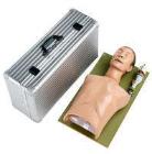
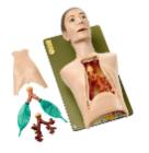
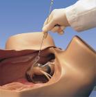
Simulator Intubation Dummy, Natural size, functional model, which has been developed in cooperation with the Federal Centre for Health Education in Cologne, makes it possible to learn how to intubate under life-like conditions, Length: 42 cm, Width: 71 cm, Height: 25 cm
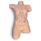
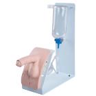
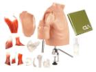
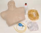
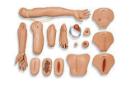
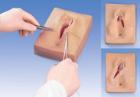
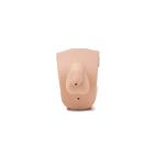
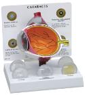
Catheterization Simulator Basic, With the male P93, Male catheterization with realistic resistance, 3 levels of adjustable narrowing of urethra, Soft and movable foreskin, Liquid outflow if catheterization is successfully carried out, Easy to clean
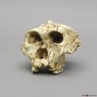
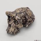
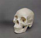
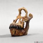
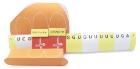
Model A. Robustus Sk-48 Cranium, 1.5-2 MYA, was discovered by Fourie in South Africa in 1950. SK-48, formerly Paranthropus crassidens, greatly increased what is known about australopithecines, Size: 15.6L x 15.1W X 12.3H (cm)
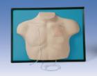
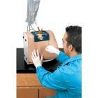
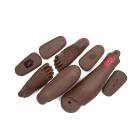
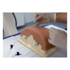
Simulator Wound Care Bandaging, For trainee nurses and doctors, students learn how to care correctly for their patients wounds and can practice a wide range of dressing and bandaging techniques. Hygiene standards and protocols can be respected.
Model Sahelanthropus Tchadensis Cranium, 6-7 MYA, was discovered by Brunet's team in 2001, Chad. Some suggest it existed near the time that hominids & apes separated on their evolutionary paths, Size: 18L x 10.2W x 10H (cm)
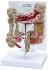
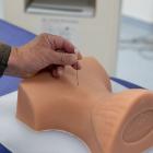
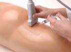
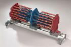
Simulator Guided Thoracic Injection, enables trainees to develop a 3D understanding of the procedures for successful spine interventions. Learn to correlate imaging with the thoracic spinal anatomy relevant to interventional pain procedures
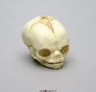
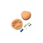
Model Fetal Human Skull 21 1/2 Weeks, Scientific Name: Homo sapiens, skull exhibits characteristics of prenatal development, 1-part skull (jaw glued to cranium), Size: 2-1/2in L x 1-3/4in W x 1-3/4in H
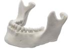
Mandible, lower jar, with 16 teeth, Model, Human Bone, Pastic, These individual bones are a great addition to you collection, used as a match for your existing Eisco disarticulated skeleton, or to allow students to compare general skeletal structure
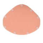
simulator, C-Section Insert Pro, for the birth simulator, P90, is ready for sectioning and tearing, The insert can be used to demonstrate and practice a C-section birth, Dimesnions: 23 x 19 x 1 cm, Weight: 0.17 kg
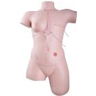
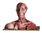
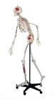
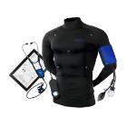
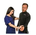
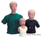
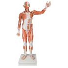
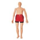
Simulator Bionic Hybrid, the suit simulates physiological conditions to test diagnostic and procedural skills. It is controlled wirelessly with easy to use software and responds in real time to diagnosis and treatment with direct feedback, Size: XXL
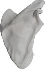
Scapula, right (shoulder blade), Model, Human Bone, Plastic, These individual bones are a great addition to you collection, used as a match for your existing Eisco disarticulated skeleton, or to allow students to compare general skeletal structure
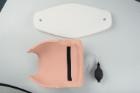
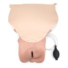
Simulator Leopold insert, The inflatable uterus set serves as a spare part for the birth simulators, Delivery contents: Inflatable uterus insert, Hand pump and tube, Sectionable upper uterine wall, Dimesnions: 46 x 27 x 13 cm, Weight: 1.15 kg
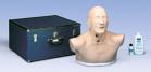
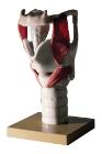
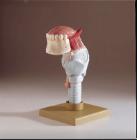
Simulator SCOPIN-Broncho-Boy II, Life-sized, in plastic, Retroflexion and left-right rotation of head for laryngoscopic, orotracheal and bronchoscopic intubation.2. Acoustic warning signal if excessive pressure exerted on upper teeth by rigid tube or laryngoscope
Model Human Female Asian Economy Skull, Features no cranial sites for musculofascial attachment, a narrow ascending mandibular ramus, smooth nasion, sharp supraorbital margins & a rounded inferior border of mandible, Size: 17.1x12x16.5cm
simulator, PPH Trainer P97 - Module, is a postpartum hemorrhage module designed to be easily integrated into the Birthing Simulator P90 PRO and BASIC, is designed to be an affordable, versatile obstetric training tool, versatile obstetric training tool
Model Sivapithecus Indicus Skull, 8.5-12.5 MYA, was discovered in 1979 by Pilbeam and Shah in Pakistan. This specimen consists of a nearly complete mandible and the left side of the face, Size: 13.4L x 11.3W x 15H (cm)
Simulator Guided Cervical Injection, enables trainees to develop a 3D understanding of the procedures for successful spine interventions. Learn to correlate imaging with the cervical spinal anatomy relevant to interventional pain procedures
Horse Skull (Equus caballus), is a great way to study the anatomy of Equus caballus. This animal skull is perfect for mammal and comparative anatomy studies