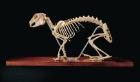
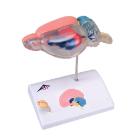
Model Rat Brain Comparative Anatomy, model shows a rat brain in approx. 6-fold enlargement. Sectioned medially, two halves, The right half of the model shows the structures of the cerebrum, cerebellum and brain stem, Dimensions : 14 x 10 x 16 cm
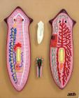
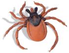
Model, Castor Bean Tick (Ixodes ricinus), Accurately detailed replica of the castor-bean tick; scale: 25:1, Dimensions: 12 x 12 x 2 cm, weight: 0.04 kg
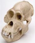
Model, Orangutan Skull (Pongo pygmaeus), Male, Replica, Primate skulls particular suitable for comparative studies, With detailed description of distinctive features, The templates for the castings were original, Dimensions: 22 x 16 x 18 cm, weight: 0.95 kg
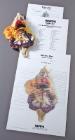
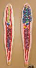
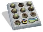
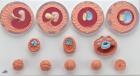
Model, Embryo Development of Common Frog, (Rana temporaria), 12 Stages, the various stages of development of an embryo, cleavage (morula and blastula), gastrulation, neurula and organogenesis enlarged 30 times, Dimensions: 37 x 36 x 13 cm, weight: 1.73 kg
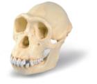
Model, Chimpanzee Skull (Pan troglodytes), Female. Replica, suitable for comparative studies, With detailed description of distinctive features, The templates for the castings were original skulls, Dimensions: 17 x 11.5 x 14 cm, weight: 0.62 kg
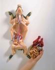
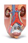
Model, Urinary System - Male - 3/4 Life Size, The right kidney of the male urinary system model is opened. This urinary system model has anatomical detail that makes it great for classroom or doctor's office, Dimensions: 10 x 18 x 26 cm, weight: 0.84 kg
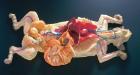
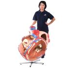
Model, Giant Heart, 8 times life size, The atria and ventricles of the heart are open to give a view of the interior, and show the accurately modeled bicuspid and major vessels adjacent to the heart, Dimensions: 100 x 90 x 70 cm, weight: 35 kg
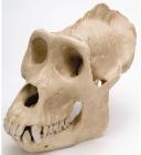
Model, Gorilla Skull (Gorilla gorilla), Male, Replica, Primate skulls particular suitable for comparative studies, With detailed description of distinctive features, The templates for the castings were original, Dimensions: 26 x 16.5 x 19.5cm, weight: 1.06kg
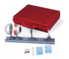
Model, Physical Eye, can be used to demonstrate the optical functions of the human eye, Half eyeball with adjustable iris diaphragm, lens holder and 2 convex lenses (f = 65 mm and 80mm), on a rod, Dimensions: 49 x 5.5 x 18 cm, Weight: 2.45 kg
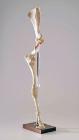
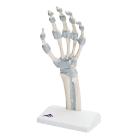
Model, Hand skeleton, with elastic ligaments, The fibrous layer of connective tissue, described in anatomy as the membrana interossea, is shown. It extends between both of these long bones, retinaculum flexorum, Dimensions: 14 x 10 x 28 cm, weight: 0.24 kg
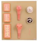
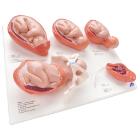
Model, Labor Stages, mounted individually bases: Fetus in womb, cervix closed, Fetus in womb, cervix open, Fetus in womb, start of head passage, Fetus in womb and pelvis, finish of head passage, Placenta in the womb, Dimensions: 40 x 31 x 13 cm, weight: 2 kg
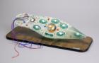
Model, Embryonic Development, 12 stages, represents the development of the human germ cells from fertilisation until the end of the 2nd month of pregnancy in 12 stages, tests for the embryological specialist field, Dimensions: 65 x 34.5 x 6 cm, weight: 0.832 kg