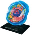
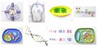
Science Animal Cell Model 4D, 6in model contains 26 detachable, hand-painted parts and a display stand, Transparent outside for seeing internal structures and super detailed parts, Also includes illustrated assembly guide, description of the anatomy, For Ages 8 plus
Horse Skull (Equus caballus), is a great way to study the anatomy of Equus caballus. This animal skull is perfect for mammal and comparative anatomy studies
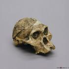
Model A.Africanus Sts 5 Mrs. Ples Cranium, 2.5 MYA, was discovered in 1947 by R. Broom and J. Robinson in Sterkfontein, Transvaal, South Africa, Size: 17.2L x 12.3W X 13.1H (cm)
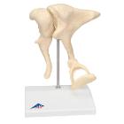
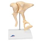
Model Ossicles Magnified 20 Times, joined to each other in the human body are located in the middle ear and are referred to as the auditory ossicles: malleus (hammer), incus (anvil) und stapes (stirrup), Weight: 0.385 kg, Dimensions : 17 x 12 x 21 cm
Model A. Africanus Sts 5 Mrs. Ples Skull, 2.5 MYA, was discovered in 1947 by R. Broom and J. Robinson in Sterkfontein, Transvaal, South Africa, Size: 17.1L x 12.1W x 16H (cm)
Model Ossicles 20X Bonelike, enlargement of original ossicles, created using micro CT, three smallest bones, joined to each other in the human body are located in the middle ear: malleus (hammer), incus (anvil) und stapes (stirrup), Dimensions : 17 x 12 x 21 cm