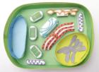
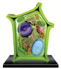
Science Plant Cell Model 4D, 6in model contains 24 detachable, hand-painted parts and a display stand, Transparent outside for seeing internal structures and super detailed parts, Also includes illustrated assembly guide, description of the anatomy, For ages 8 Plus
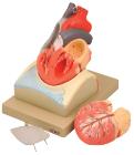
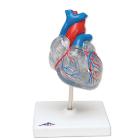
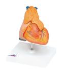
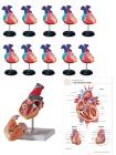
Enlarged, sectioned so that both ventricles and atria open to expose the valves. Large blood vessels near the heart and musculature of the heart are shown. Separates into 4 parts. On base.
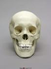
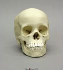
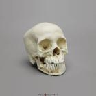
Model Human Adolescent Skull, features of race are of Asian, the totality of features is most with those of slightly gracile young male who has not yet fully developed his sexual characteristics & might be of a slightly robust female, Size: 19x14.2x20.1cm
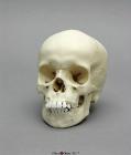
Model Human Child Skull, 13-year-old, With exception of wisdom teeth, all permanent teeth are fully erupted & no deciduous dentition remains, apices of canines, premolars & second molars are approximately one-half to two-thirds closed, Size: 7x5x7in
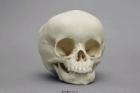
Model Human Child Skull 15-month-old, contains 12 teeth in upper & lower jaws, anterior & posterior intra-occipital sutures are not fused, anterior fontanelle is open, posterior, sphenoidal & mastoidal fontanelle are closed, Size: 15.3x12.1X12cm
Model Human Child Skull, root apices of permanent incisors & first molars are almost completely formed, spheno-occipital synchondrosis is open, anterior intra-occipital suture is closed, 2-part skull (jaw connected with hardware), Size: 18x12.6x17.1cm
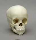
Model Human Child Skull, 10 teeth in maxillary & mandibular arcade, apices are formed on primary incisors, no visible tooth buds present at apices of primary molar, Crown formation on permanent incisors & first molar is 2/3 complete, Size: 15.9x11.5x14.3cm
Model Human Child Skull with Dentition Exposed, right side of maxilla & jaw bone has been cut away, Two of the teeth are only visible after part of mandibular cortical bone has been removed, 2-part skull (separate cranium & jaw), Size: 16.4x12x15cm
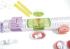
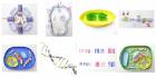
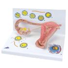
Model Stages Of Fertilisation, Dimensions: 35x21x20 cm, begin the growth into an embryo, The various stages are shown in larger-than-life model form in an ovary, fallopian tube and womb. An even more enlarged illustration of each is also printed on the base