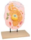
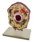
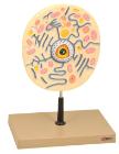
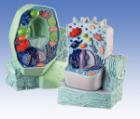
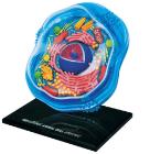
Three dimensional model showing electron microscopic structure. Organs like nucleus, Nucleolus, endoplasmic reticulum, mitochondria, ribosomes respectively polysomes and golgi apparatus. Showing centrioles, lysosomes and vacuoles.
Enlarged 20,000 times. The model shows delicate structure of an animal cell. Features organelles nucleus, endoplasmic reticulum, mitochondria, ribosomes respectively polysomic and Golgi apparatus. Also showing centrioles, lysosomes and fat vacuoles.
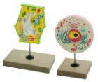
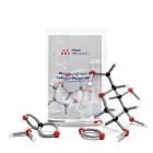
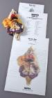
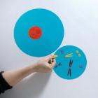
Photosynthesis and Respiration Model Set, Fundamental processes in the flow of energy, Students build glucose and oxygen molecules at the giant chloroplast, Move the molecules to the mitochondria and reassemble them into carbon dioxide and water molecule
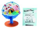
Science Animal Cell Model 4D, 6in model contains 26 detachable, hand-painted parts and a display stand, Transparent outside for seeing internal structures and super detailed parts, Also includes illustrated assembly guide, description of the anatomy, For Ages 8 plus
Science BioSigns Animal Cell Model, Hands-on interactive 14-piece model teaches about cell membranes and mitochondria, Provides a magnified and cross-sectioned detailing of an Animal Cell, Includes Guide
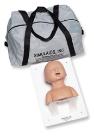
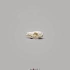
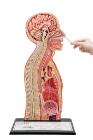
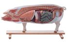
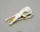
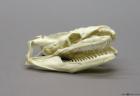
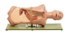
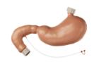
Simulator OGI Phantom, For training of endoscopy of upper gastrointestinal tract, for catheterisation of papilla vateri by retrograde instillation of contrast medium into pancreatic duct system (Natural size, made of special plastic), Esophagoscopy, gastroscopy & bulboscopy
Simulator Stomach and Duodenum, In natural size, made of special plastic. For training of endoscopy according to Prof. Dr. M. Classen and H. Ruppin, M.D, Weight: 1,3 kg, Length: 27 cm, Width: 36 cm, Height: 12 cm