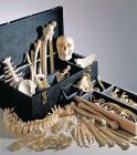
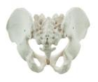
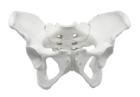
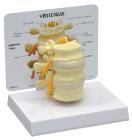
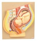
It Illustrate the morphological distinctions between male and female pelvic structures. Each pelvis includes the left and right innominates with pubic symphysis, 4th and 5th lumbar vertebrae with intervertebral discs, the sacrum and coccyx.
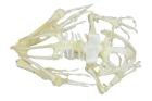
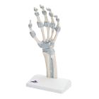
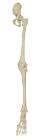
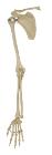
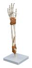
Model, Hand skeleton, with elastic ligaments, The fibrous layer of connective tissue, described in anatomy as the membrana interossea, is shown. It extends between both of these long bones, retinaculum flexorum, Dimensions: 14 x 10 x 28 cm, weight: 0.24 kg
Illustrate the morphological distinctions between male and female pelvic structures. Each pelvis includes the left and right innominates with pubic symphysis, 4th and 5th lumbar vertebrae with intervertebral discs, the sacrum and coccyx.
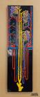
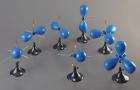
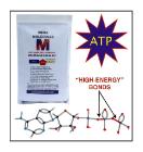
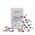
ATP and Cell energy Model Set, Includes: Carbon/Hydrogen/Oxygen/Nitrogen/Phosphorus atoms, Red links for high energy bonds, Gray links for single/double bonds, Illustrate that metabolic processes use ATP as an energy source/convert it back into inorganic phosphate/ADP
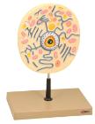
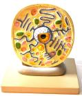
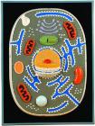
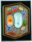
Plant Cell Model, shows electron microscopic detail of a typical plant cell, It shows cellular structures, vacuoles, nuclear structure, plastids, mitochondria and more, It is mounted on a base and numbered with a key card
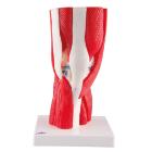
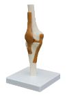
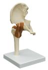
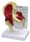
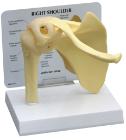
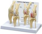
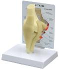
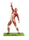
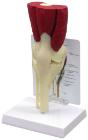
Model Knee Joint 12-Part, with removable joints is part of a high quality series of models, manufactured to precise anatomical correctness with realistic colors of the joint, bone and muscles, extremely durable, nonhazardous material of standards, Dimensions : 33x17x17 cm
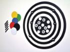
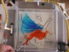
Photosynthesis and Respiration Model Set, Fundamental processes in the flow of energy, Students build glucose and oxygen molecules at the giant chloroplast, Move the molecules to the mitochondria and reassemble them into carbon dioxide and water molecule
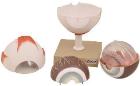
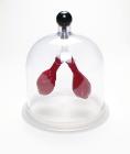
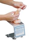
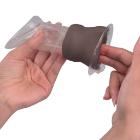
Model Female Condom Dark Skin, Dimensions: 12 cm, shows the labia and vagina up to the cervix in a simplified representation for didactic reasons, and is used for demonstrating and learning the insertion of a female condom
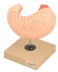
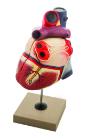
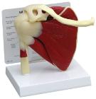
Life size model dissectible in 2 parts. The anterior heart wall can be removed to show the left and right ventricles and atria as well as the tricuspid, pulmonary, mitral and aortic valves. Mounted on base.
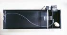
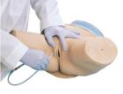
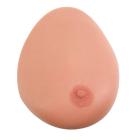
Model Single Breast, With Benign Tumor, Dimensions: 25.4 x 17.8 x 17.8 cm, made of 3B SKINlike* silicone with simulated benign tumour for the demonstration of ultrasonic B-image mode with Ultrasonic Echoscope GS200
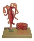
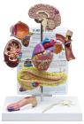
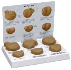
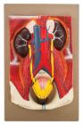
Model Human Urinary Organs, Weight (lb):2.6, Length (in):13.0,Width (in): 9.1, Height (in): 4.3,Natural Size separates into 3 parts. Kidneys ureters, adrenal glands, bladder with prostate and major blood vessels