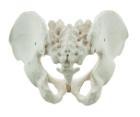
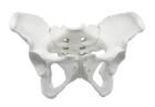
It Illustrate the morphological distinctions between male and female pelvic structures. Each pelvis includes the left and right innominates with pubic symphysis, 4th and 5th lumbar vertebrae with intervertebral discs, the sacrum and coccyx.
Illustrate the morphological distinctions between male and female pelvic structures. Each pelvis includes the left and right innominates with pubic symphysis, 4th and 5th lumbar vertebrae with intervertebral discs, the sacrum and coccyx.
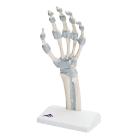
Model, Hand skeleton, with elastic ligaments, The fibrous layer of connective tissue, described in anatomy as the membrana interossea, is shown. It extends between both of these long bones, retinaculum flexorum, Dimensions: 14 x 10 x 28 cm, weight: 0.24 kg
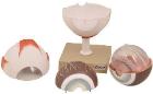
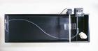
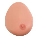
Model Single Breast, With Benign Tumor, Dimensions: 25.4 x 17.8 x 17.8 cm, made of 3B SKINlike* silicone with simulated benign tumour for the demonstration of ultrasonic B-image mode with Ultrasonic Echoscope GS200
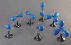
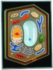
ATP and Cell energy Model Set, Includes: Carbon/Hydrogen/Oxygen/Nitrogen/Phosphorus atoms, Red links for high energy bonds, Gray links for single/double bonds, Illustrate that metabolic processes use ATP as an energy source/convert it back into inorganic phosphate/ADP