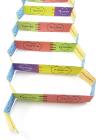
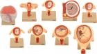
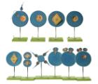
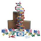
Poisoned Appreciation Model Set, Students explore the chemical properties/structural formulas of reducing sugars, starch, protein, fat, build a series of molecular models and analyze the results of chemical tests to determine which food at the luncheon was poisoned
Model Ebola Virus 10000X, represents the virus magnified approximately 100000 times in its unique shape, surface shows the viral membrane with Glycoproteins, A cut-away at one end exposes internal structures major and minor matrix proteins polymerase protein and Ebola RNA
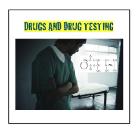
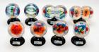
Drug testing model Set, Students act as members of a hospital administration who are responsible for developing a substance-abuse prevention/drug testing program, Students will explore the chemical and physical properties of five illicit drugs and build a model for each drug
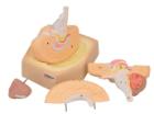
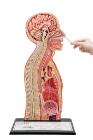
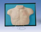
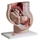
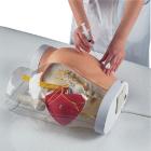
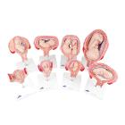
Set of nine models, showing the following stages. 1. Embryo 6 days old 2. 1st month of gestation. 3. Uterus with embryo in 3rd month of gestation.4. Uterus with fetus, in 4th month. 5. Uterus with fetus, placenta and umbilical cord.6. 5th month. 7. 7th month pregnancy.
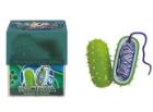
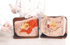
Bacteria Model, Hands-on interactive model provides a magnified and cross-sectioned detailing of Bacteria, Able to be separated, with both halves filled with a cross section of genetic material and flagellum, With Guide
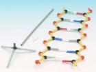
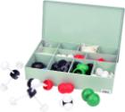
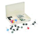
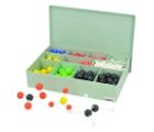
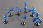
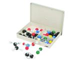
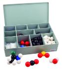
Model Atomic Set - Economy, Senior, Idealy suited for advance level courses, Used for modeling three dimensional simple or complex molecular structures of organic and inorganic compounds, set consists of 150 connecting lugs and 370balls in different sizes and colors
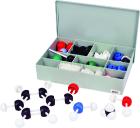
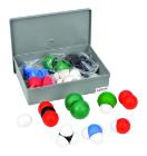
Molecular Modelling set, Basic Inorganic and Organic Chemistry, Hard plastic case, Standard size, Consists of 54 balls of various colours and 24 links, 64 Pieces, Carbon-12, Hydrogen-24, Oxygen-6, Nitrogen-6, Chlorine-2, Sulphur-2, Bromine-1, Iodine-1
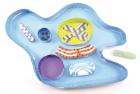
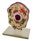
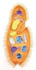
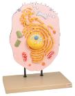
Enlarged 20,000 times. The model shows delicate structure of an animal cell. Features organelles nucleus, endoplasmic reticulum, mitochondria, ribosomes respectively polysomic and Golgi apparatus. Also showing centrioles, lysosomes and fat vacuoles.
Three dimensional model showing electron microscopic structure. Organs like nucleus, Nucleolus, endoplasmic reticulum, mitochondria, ribosomes respectively polysomes and golgi apparatus. Showing centrioles, lysosomes and vacuoles.
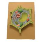
This plant cell model is separated in 4 parts. Showing details of cell wall and inner details of cell wall, nucleus is separated and defined in a 3-D way cut section of chloroplast is shown.
Virus Model, Hands-on interactive model provides a magnified and cross-sectioned detailing of Bacteria, Able to be separated, with both halves filled with a cross section of genetic material and flagellum, With Guide