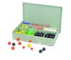
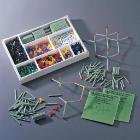
Model Atomic Set - Economy, Senior, Idealy suited for advance level courses, Used for modeling three dimensional simple or complex molecular structures of organic and inorganic compounds, set consists of 150 connecting lugs and 370balls in different sizes and colors
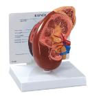
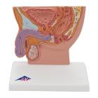
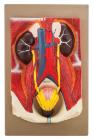
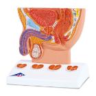
Model Human Urinary Organs, Weight (lb):2.6, Length (in):13.0,Width (in): 9.1, Height (in): 4.3,Natural Size separates into 3 parts. Kidneys ureters, adrenal glands, bladder with prostate and major blood vessels
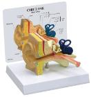
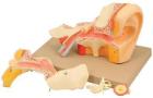
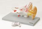
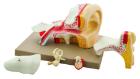
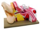
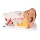
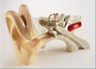
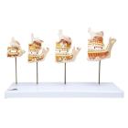
Enlarged approximately 4 times. The petros portion of the temporal bone section of the auditory canal are removable, with labyrinth can be taken out and opened. The tympanic membrane with malleus and incus can be removed in 5 parts.
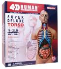
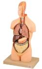
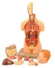
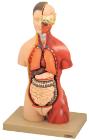
Human Anatomy Deluxe Torso 4D, 15in model contains 54 detachable, hand-painted medical education-quality organs and parts and a display stand, Also includes illustrated assembly guide and description of the anatomy, Offer a unique puzzle challenge, For ages 14 plus
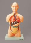
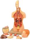
Half size African torso, can be dissected into 9 parts. Half life-size can be quickly disassembled this torso, by removing lungs and heart-2 parts, stomach, liver and gall bladder, intestine 2 parts. Includes key card
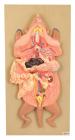
Showing the harmful effects of hypertension on the most susceptible organs. It consists of scaled down depictions of brain, eye, 2-part heart, 2-part kidney, enlarged artery. All of the organs can be rotated or removed for closer viewing.
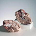
Half size torso, sexless. Head is dissectible in 3 parts showing half of the brain can be taken out, showing the cranial nerves and the important inteRNAl parts of the skull.
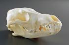
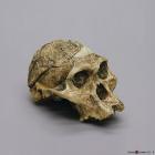
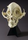
Model A.Africanus Sts 5 Mrs. Ples Cranium, 2.5 MYA, was discovered in 1947 by R. Broom and J. Robinson in Sterkfontein, Transvaal, South Africa, Size: 17.2L x 12.3W X 13.1H (cm)
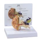
Model Ossicles Magnified 20 Times, joined to each other in the human body are located in the middle ear and are referred to as the auditory ossicles: malleus (hammer), incus (anvil) und stapes (stirrup), Weight: 0.385 kg, Dimensions : 17 x 12 x 21 cm
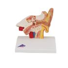
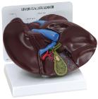
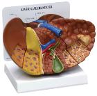
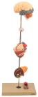
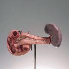
Rear Organs Of The Upper Abdomen, The upper abdomen organ model shows the duodenum (partially opened), gall bladder (opened) and bile ducts (opened), the pancreas (revealing large ducts), the spleen and the surrounding vessels in natural size
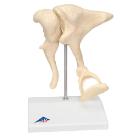
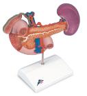
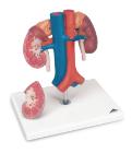
Kidneys with Vessels 2-Part. This kidney model shows the kidneys with suprarenal glands, the outgoing ureters, the renal vessels and the large vessels situated close to the kidneys in natural size.
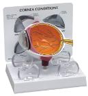
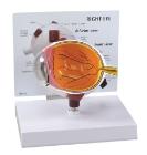
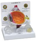
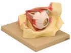
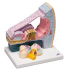
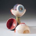
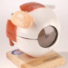
Model shows both sides of an eye, enlarged 5 x. One side of the model shows the eye socket with a sagittal cutaway and the background to the eye and the electron microscopic fine structure of the retina are shown separately.
Model A. Africanus Sts 5 Mrs. Ples Skull, 2.5 MYA, was discovered in 1947 by R. Broom and J. Robinson in Sterkfontein, Transvaal, South Africa, Size: 17.1L x 12.1W x 16H (cm)
Model Organ Of Corti, three dimensional section through the organ of Corti, the site of the sense of hearing in the inner ear in humans, detailed representation of the individual cellular components and membranes, exact location of the organ, Dimensions : 26 x 19 x 26 cm
Model Ossicles 20X Bonelike, enlargement of original ossicles, created using micro CT, three smallest bones, joined to each other in the human body are located in the middle ear: malleus (hammer), incus (anvil) und stapes (stirrup), Dimensions : 17 x 12 x 21 cm
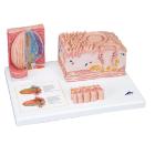
Model Microanatomy* Tongue, specific parts of the tongue, macroscopic view of the tongue in life size (dorsal view) and microscopic views of the various papillae of the tongue (10-20x life size) and of a taste bud ( 450x life size), Dimensions : 14.5 x 32.5 x 20 cm