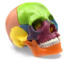
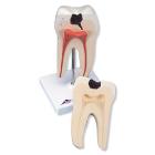
Didactic Painted Mini Skull Model, Reduced size (1/2 natural size), anatomically correct replica, Realistic details, texture and bony landmarks make this model ideal for educational demonstration, Multicolored pieces for easier identification, 15 Pieces
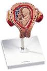
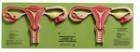
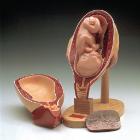
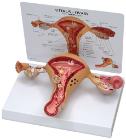
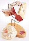
Model, Embryo, 3 month, The embryo model shows an embryo at the stage of the first month of pregnancy, Ideal for medical training and as an educational tool for pregnant women, Important anatomical structures are labeled
Model, Embryo, 1 month, The embryo model shows an embryo at the stage of the first month of pregnancy, Ideal for medical training and as an educational tool for pregnant women, Important anatomical structures are labeled, weight: 0.33 kg
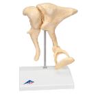
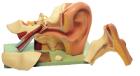
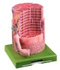
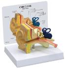
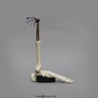
Model Ossicles 20X Bonelike, enlargement of original ossicles, created using micro CT, three smallest bones, joined to each other in the human body are located in the middle ear: malleus (hammer), incus (anvil) und stapes (stirrup), Dimensions : 17 x 12 x 21 cm
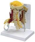
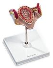
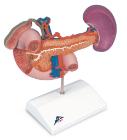
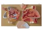
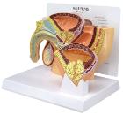
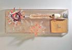
Rear Organs Of The Upper Abdomen, The upper abdomen organ model shows the duodenum (partially opened), gall bladder (opened) and bile ducts (opened), the pancreas (revealing large ducts), the spleen and the surrounding vessels in natural size
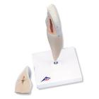
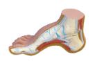
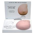
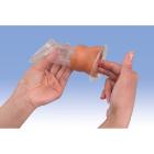
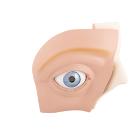
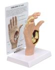
Model Female Condom Dark Skin, white skin tone, Dimensions: 12 cm, shows the labia and vagina up to the cervix in a simplified representation for didactic reasons, and is used for demonstrating and learning the insertion of a female condom
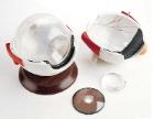
Model, Eye, 5 times full-size, 12 part, Two halves of the sclera, Optic nerve, M. Rectus superior, M. Rectus lateralis, Cornea half, Eye lens, Lachrymal system, Vitreous humour, Tear gland, Associated structures, Dimensions: 33 x 30 x 38 cm, weight: 4.382 kg