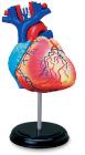
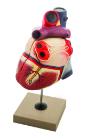
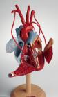
Enlarged. Sectioned so that both ventricles and atria open to expose the valves. Large blood vessels near the heart and musculature of the heart are shown. Separates into 7 parts.
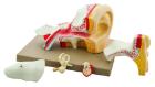
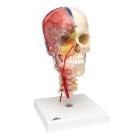
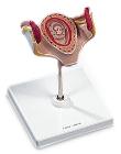
Enlarged approximately 4 times. The petros portion of the temporal bone section of the auditory canal are removable, with labyrinth can be taken out and opened. The tympanic membrane with malleus and incus can be removed in 5 parts.
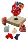
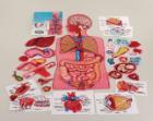
4D Anatomy Human Heart Model, 4.5in model contains 32 detachable, hand-painted medical education-quality organs and parts and a display stand, Also includes illustrated assembly guide and description of the anatomy, Has unique puzzle challenge, For ages 8plus
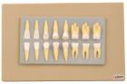
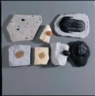
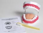
Enlarged approximately 2 times, set of 16 teeth cast in break resistant material having accurate anatomical details. Complete set as in half of upper & lower jaw
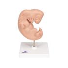
Model, Embryo, 3 month, The embryo model shows an embryo at the stage of the first month of pregnancy, Ideal for medical training and as an educational tool for pregnant women, Important anatomical structures are labeled
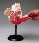
Life size model dissectible in 2 parts. The anterior heart wall can be removed to show the left and right ventricles and atria as well as the tricuspid, pulmonary, mitral and aortic valves. Mounted on base.
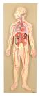
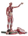
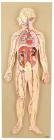
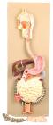
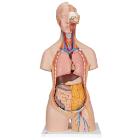
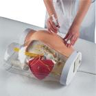
This half-size relief model helps in understand the circulation of blood in body through brain, heart, lung, liver, spleen, kidneys and partial skeleton. Mounted on base. Numbered with key card.
Lower incisor with removable half of crown, lower canine separates in two longitudinally, lower molar with one root is one-piece and two roots separate into three parts, upper molar with three roots separates into three parts.
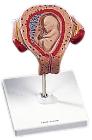
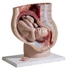
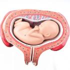
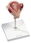
Model, month 5, dorsal position, shows a fetus in the fifth month in the dorsal position in the uterus, All important anatomical structures are labeled, weight: 0.54 kg
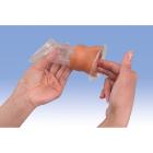
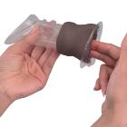
Model Female Condom Dark Skin, white skin tone, Dimensions: 12 cm, shows the labia and vagina up to the cervix in a simplified representation for didactic reasons, and is used for demonstrating and learning the insertion of a female condom
Model Female Condom Dark Skin, Dimensions: 12 cm, shows the labia and vagina up to the cervix in a simplified representation for didactic reasons, and is used for demonstrating and learning the insertion of a female condom
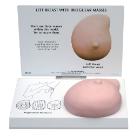
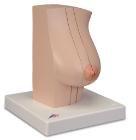
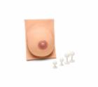
Model Of The Female Breast, Medially divided into 2 halves, held together with magnets, Healthy milk-giving breath tissue on the cut surface of the external half, Breast gland inflammation (mastitis) on the cut surface of the inner half
Model, Embryo, 2 month, The embryo model shows an embryo at the stage of the first month of pregnancy, Ideal for medical training and as an educational tool for pregnant women, Important anatomical structures are labeled, weight: 0.38 kg
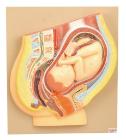
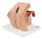
Model For Gynecological Patient Education, This unique gynecological training model is ideal for demonstration purposes and for realistic insertion of female barrier contraceptive devices, which are placed in the vaginal/cervical area.
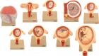
Set of nine models, showing the following stages. 1. Embryo 6 days old 2. 1st month of gestation. 3. Uterus with embryo in 3rd month of gestation.4. Uterus with fetus, in 4th month. 5. Uterus with fetus, placenta and umbilical cord.6. 5th month. 7. 7th month pregnancy.
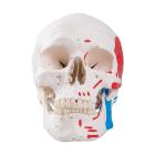
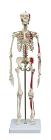
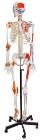
Human Skeleton Model, with Ligament Details, Painted Muscle Origins and Insertions, Flexible Spine and Joints - Rod Mount with Rolling Base, Numbered keycard, anatomical chart and dust cover included
Model, Embryo, 1 month, The embryo model shows an embryo at the stage of the first month of pregnancy, Ideal for medical training and as an educational tool for pregnant women, Important anatomical structures are labeled, weight: 0.33 kg
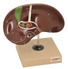
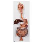
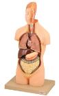
Life size model showing nose, mouth cavity and pharynx, liver with gall bladder, pancreas and spleen. Transverse colon and front stomach wall are removable. Mounted on base. Numbered with Key Card
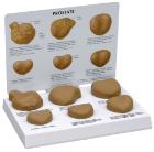
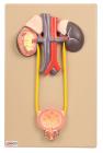
Natural size. Kidneys, ureters, adrenal glands and bladder with prostate as well as the large abdominal right kidney sectioned to show all anatomical details.
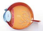
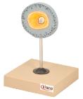
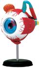
Human Anatomy Eyeball Model 4D, 4.5in model contains 35 detachable, hand-painted medical education-quality organs and parts and a display stand, Also includes illustrated assembly guide and description of the anatomy, Offer a unique puzzle challenge, For ages 8 plus