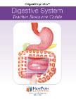
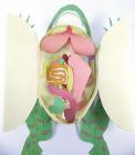
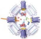
Human Anatomy Transparent Torso Model 4D, 8in model contains 37 detachable, hand-painted medical education-quality organs and parts and a display stand, Also includes illustrated assembly guide and description of the anatomy, For ages 8 plus
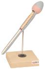
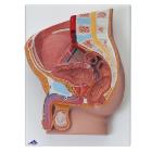
Model For Gynecological Patient Education, This unique gynecological training model is ideal for demonstration purposes and for realistic insertion of female barrier contraceptive devices, which are placed in the vaginal/cervical area.
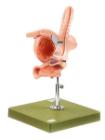
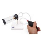
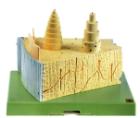
Simulator Catheterisation-Model, male, Ideal for demonstrating disposable & ballon catheters as well as suprapubic aspiration. Can be dismantled into two parts, natural size, made of special plastic. On a stand with green base, LxWXH: 18 X 18 X 30 cm
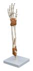
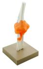
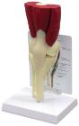
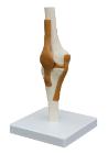
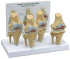
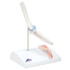
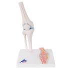
Model of human elbow joint. Mounted on base. Features upper half of radius and ulna, lower half of humerus and the ligaments of the elbow joint, to show hinge joint, Numbered with English Key Card, Size 17x 22 x 22 cm approx, Weight 250 g
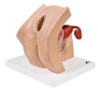
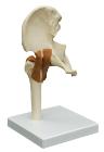
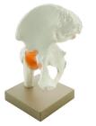
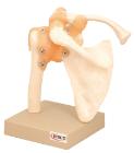
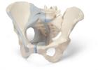
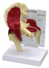
Model of human hip joint. With simulated pubofemoral ligament, Mounted on base, represents the upper femur and pelvic bone and the way that they move, Numbered with English Key Card, Size 22 x 12 x 25 cm approx. Weight 800 g approx
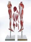
Mini-Skeleton with Flexible Spine, flexible spine to demonstrate natural movement of spine and thorax. Ideal for the chiropractor and orthopedic surgeon. Bones of the skeleton are individually represented - results in full articulation and movements of joints
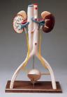
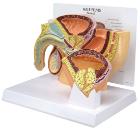
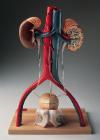
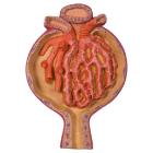
Renal Corpuscle Kidney 700 Times Full-Size, The Malpighian Corpuscle kidney model shows an open Malpighian corpuscle with glomerulus and Bowman's capsule
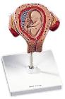
Model, Embryo, 3 month, The embryo model shows an embryo at the stage of the first month of pregnancy, Ideal for medical training and as an educational tool for pregnant women, Important anatomical structures are labeled
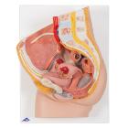
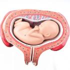
Model, month 5, dorsal position, shows a fetus in the fifth month in the dorsal position in the uterus, All important anatomical structures are labeled, weight: 0.54 kg