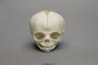
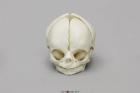
Model Fetal Human Skull 30 Weeks, Scientific Name: Homo sapiens, skull exhibits characteristics of prenatal development, 1-part skull (jaw glued to cranium), Size: 3in L x 2-1/2in W x 2-1/2in H
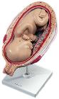
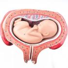
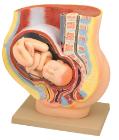
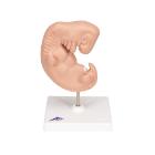
Model, Fetus, month 7, The model shows a fetus in the seventh month in the uterus, All important anatomical structures are labeled, Dimensions: 15 x 32 x 27 cm, weight: 0.86 kg
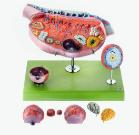
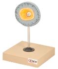
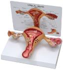
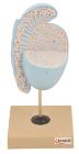
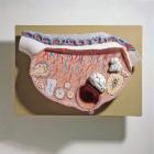
Model of the Ovary, Enlarged approximately 10 times, in SOMSO-Plast* Horizontal section parallel to the mesovarian margin with presentation of the follicles in different maturation phases, corpus rubrum, luteum and albicans, Separates into 13 parts. Mounted
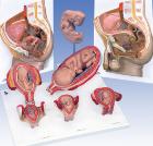
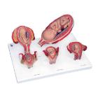
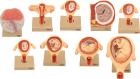
Set of nine models, showing the following stages. 1. Embryo 6 days old 2. 1st month of gestation. 3. Uterus with embryo in 3rd month of gestation.4. Uterus with fetus, in 4th month. 5. Uterus with fetus, placenta and umbilical cord.6. 5th month. 7. 7th month pregnancy.
Model, month 5, dorsal position, shows a fetus in the fifth month in the dorsal position in the uterus, All important anatomical structures are labeled, weight: 0.54 kg
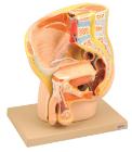
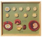
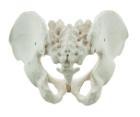
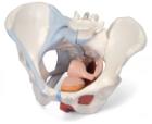
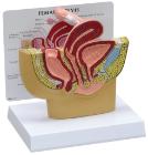
It Illustrate the morphological distinctions between male and female pelvic structures. Each pelvis includes the left and right innominates with pubic symphysis, 4th and 5th lumbar vertebrae with intervertebral discs, the sacrum and coccyx.
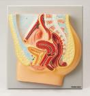
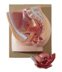
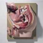
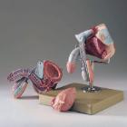
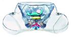
Female Pelvis with Ligaments 4 Part, This life size four part model of a female pelvis represents detailed information about the topography of bones, ligaments, pelvic floor muscles and female pelvic organs.
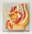
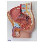
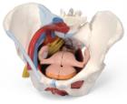
Female Pelvis with Ligaments 6 Part, This life size six part model of a female pelvis represents detailed information about the topography of bones, ligaments, vessels, nerves, pelvic floor muscles and female genital organs.
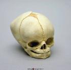
Model Fetal Human Skull 40 1/2 Weeks, Full term, skull exhibits characteristics of prenatal development, Scientific Name: Homo sapiens, 1-part skull (jaw glued to cranium), Size: 11.9L x 9.1W x 8.5H (cm)
Model Fetal Human Skull 29 Weeks, Scientific Name: Homo sapiens, skull exhibits characteristics of prenatal development, 1-part skull (jaw glued to cranium), Size: 3in L x 2-1/4in W x 2-1/2in H
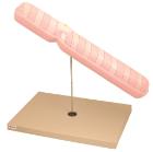
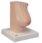
Model Of The Female Breast, Medially divided into 2 halves, held together with magnets, Healthy milk-giving breath tissue on the cut surface of the external half, Breast gland inflammation (mastitis) on the cut surface of the inner half