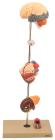
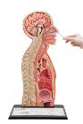
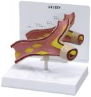
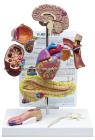
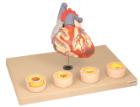
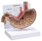
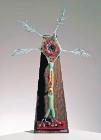
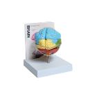
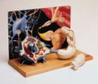
Showing the harmful effects of hypertension on the most susceptible organs. It consists of scaled down depictions of brain, eye, 2-part heart, 2-part kidney, enlarged artery. All of the organs can be rotated or removed for closer viewing.
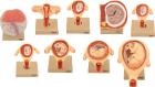
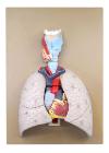
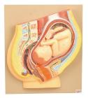
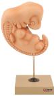
Set of nine models, showing the following stages. 1. Embryo 6 days old 2. 1st month of gestation. 3. Uterus with embryo in 3rd month of gestation.4. Uterus with fetus, in 4th month. 5. Uterus with fetus, placenta and umbilical cord.6. 5th month. 7. 7th month pregnancy.
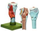
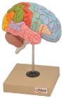
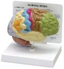
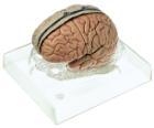
Life size, half of human brain. Students can study regions within the cerebral cortex are allied to certain functions. Impulses from the sensory organs, the skeletal muscles, skin and joints all travel to areas specialized in interpreting the information
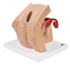
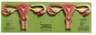
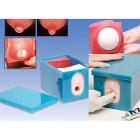
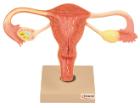
Model For Gynecological Patient Education, This unique gynecological training model is ideal for demonstration purposes and for realistic insertion of female barrier contraceptive devices, which are placed in the vaginal/cervical area.
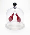
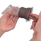
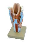
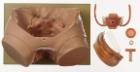
Model Female Condom Dark Skin, Dimensions: 12 cm, shows the labia and vagina up to the cervix in a simplified representation for didactic reasons, and is used for demonstrating and learning the insertion of a female condom
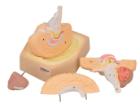
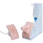
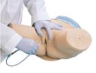
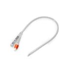
BASIC catheterization trainer set, both male and female bladder catheterization procedures can be realistically demonstrated, practiced and assessed. The easy-to-change genital inserted into the holder and held in place with magnets, with Both inserts
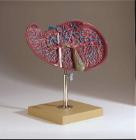
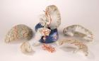
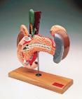
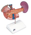
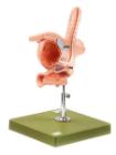
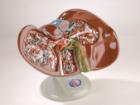
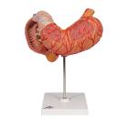
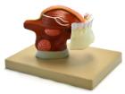
Rear Organs Of The Upper Abdomen, The upper abdomen organ model shows the duodenum (partially opened), gall bladder (opened) and bile ducts (opened), the pancreas (revealing large ducts), the spleen and the surrounding vessels in natural size
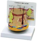
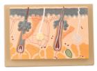
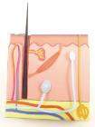
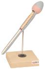
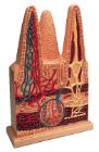
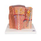
Enlarged 70 Times, full size, shows section through three layers of the hair covered skin of the head. Also shows hair follicles with sebaceous, sweat glands, receptors, nerves and vessels.
Simulator Catheterisation-Model, male, Ideal for demonstrating disposable & ballon catheters as well as suprapubic aspiration. Can be dismantled into two parts, natural size, made of special plastic. On a stand with green base, LxWXH: 18 X 18 X 30 cm
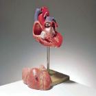
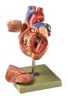
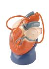
Sectioned through the ventricles and auricles. The bicuspid and tricuspid semilunar and sigmoid valves are shown. Separates into 3 parts. Mounted on base
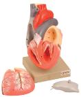
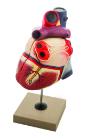
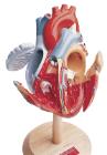
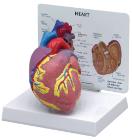
Heart dissected in two parts shows the thickened ventricle walls and cardiac valves. Four individual blood vessel model show coronary artery disease/ atherosclerosis and progression in the blood vessel, myocardial infarction and damage.
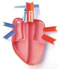
Life size model dissectible in 2 parts. The anterior heart wall can be removed to show the left and right ventricles and atria as well as the tricuspid, pulmonary, mitral and aortic valves. Mounted on base.
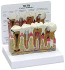
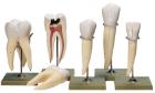
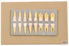
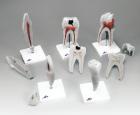
Enlarged approximately 2 times, set of 16 teeth cast in break resistant material having accurate anatomical details. Complete set as in half of upper & lower jaw
Model-Heart two parts, 100cm tall, Communicate the basics of heart anatomy to beginning biology students, Colorful and durable, features both external and internal components, including valves, two-piece model is mounted on a stand and can be removed for up-close study
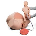
Birthing Simulator PRO, developed for the skill training in normal deliveries, in complicated deliveries, in obstetric emergencies, Obstetric simulation has proven successful to enhance the training of delivery skill, following of protocols and reaction in emergency situation
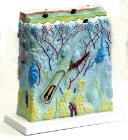
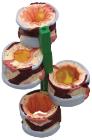
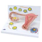
Model Stages Of Fertilisation, Dimensions: 35x21x20 cm, begin the growth into an embryo, The various stages are shown in larger-than-life model form in an ovary, fallopian tube and womb. An even more enlarged illustration of each is also printed on the base