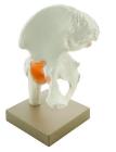
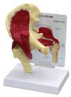
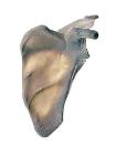
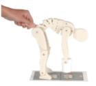
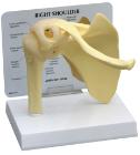
Model of human hip joint. With simulated pubofemoral ligament, Mounted on base, represents the upper femur and pelvic bone and the way that they move, Numbered with English Key Card, Size 22 x 12 x 25 cm approx. Weight 800 g approx
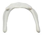
Hyoid bone (lingual bone), Model, Human Bone, Plastic, These individual bones are a great addition to you collection, used as a match for your existing Eisco disarticulated skeleton, or to allow students to compare general skeletal structure
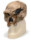
Model, Replica Homo steinheimnensis Skull, (Berkhemer, 1936), is a detailed casting from Berkhemer's reconstruction (1936, skull with no jawbone), The Steinheim replica was modeled after an original skull, Dimensions: 19 x 12.5 x 21.5 cm, weight: 1.14 kg
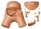
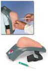
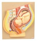
Simulator Model for pelviscopic operations, model consists of a rigid plastic mass and corresponds to the actual size of a normal woman, Many operations are made possible by number of openings, designed mainly for simulation of all pelviscopic operations
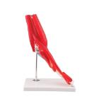
Model Elbow Joint 8 Parts, great tool for student and patient education, part of a high quality series of muscle models, and has been manufactured to replicate the anatomy of the human elbow joint in detail, colors used, Weight: 1.74 kg, Dimensions : 25 x 41 x 25 cm
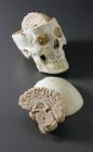
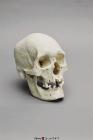
Human Male Polynesian Skull, sagittal keel, parietal bossing, the broad, prominent basiooccipital, and the rocker jaw, rocker jaw is curved along the inferior surface of the mandible, 2-part skull (separate cranium & jaw), Size: 20.7x14.2x19.6cm
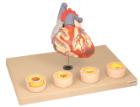
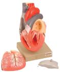
Heart dissected in two parts shows the thickened ventricle walls and cardiac valves. Four individual blood vessel model show coronary artery disease/ atherosclerosis and progression in the blood vessel, myocardial infarction and damage.
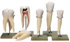
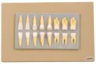
Enlarged approximately 2 times, set of 16 teeth cast in break resistant material having accurate anatomical details. Complete set as in half of upper & lower jaw
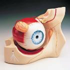
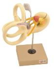
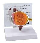
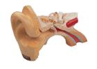
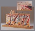
Model shows both sides of an eye, enlarged 5 x. One side of the model shows the eye socket with a sagittal cutaway and the background to the eye and the electron microscopic fine structure of the retina are shown separately.
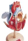
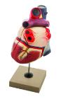
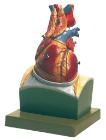
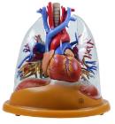
Life size model dissectible in 2 parts. The anterior heart wall can be removed to show the left and right ventricles and atria as well as the tricuspid, pulmonary, mitral and aortic valves. Mounted on base.
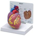
Model-Heart two parts, 100cm tall, Communicate the basics of heart anatomy to beginning biology students, Colorful and durable, features both external and internal components, including valves, two-piece model is mounted on a stand and can be removed for up-close study
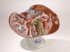
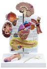
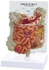
Life size model showing nose, mouth cavity and pharynx, liver with gall bladder, pancreas and spleen. Transverse colon and front stomach wall are removable. Mounted on base. Numbered with Key Card
Sectioned through the ventricles and auricles. The bicuspid and tricuspid semilunar and sigmoid valves are shown. Separates into 3 parts. Mounted on base